Calls for Ukraine
Calls for Europe
Calls for USA

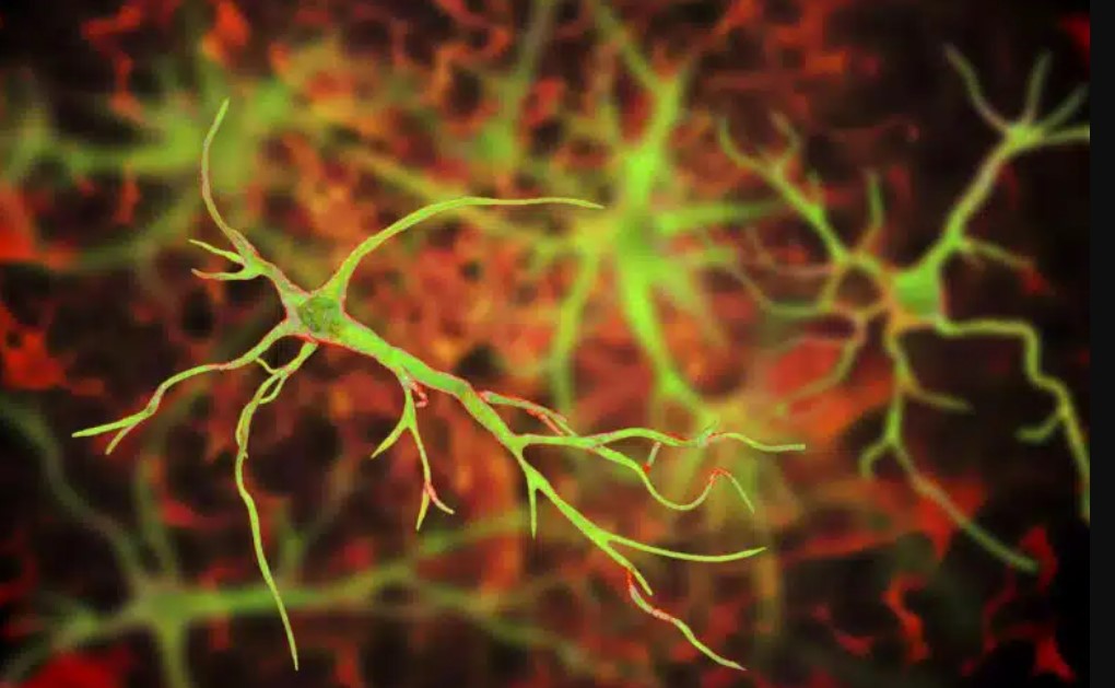
Astrocytes (cells named for their star shape) are a type of brain cell as numerous as neurons in the central nervous system, but little is known about their role in brain health and disease.
We do know that many neurological diseases are caused by or result in cell loss in the central nervous system. Some diseases result from the loss of specific cells, for example, in amyotrophic lateral sclerosis (ALS) there is a loss of motor neurons, in Parkinson’s disease there is a loss of dopaminergic neurons, and in Huntington’s disease there is a loss of GABAergic neurons.
Although many brain diseases are characterized by the loss of specific cells, the common link among all these diseases is the loss of astrocytes.
Evidence to date suggests that astrocytes are involved in basic brain functions, including homeostasis and modulation of neural networks, which are essential for everyday cognition. Healthy astrocytes are essential for brain function, and therefore finding strategies to cure or replace damaged astrocytes may help treat many neurological diseases.
Cell replacement therapy involves transplanting functional cells to patients. In recent years, the area has seen interesting developments in astrocyte transplantation in animal models of disease, and one approach has even moved into early clinical trials for patients with ALS. Despite promising results, the success rate of treatment varies from study to study.
A recent study published in The Journal of Neuroscience examined how transplanted astrocytes integrate into the recipient’s central nervous system. The types of transplanted astrocytes, timing of treatment, and transplantation methods were studied.
First, the scientists prepared cultures of astrocytes in petri dishes, extracting immature astrocytes from the cerebral cortex of newborn mice and increasing the cell population, and then transplanted them into the brains of other mice.
It turned out that the transplanted astrocytes could normally develop and integrate into the recipient’s brain, and function similarly to their own astrocytes (with minor differences).
The ability of astrocytes to perceive signals and exchange information in the brain is accomplished by molecules such as receptors and ion channels located on their cell surface. Transplanted astrocytes show a comparable number of these receptors and channels and are of similar size and complexity compared to the recipients’ own astrocytes.
The transplanted astrocytes take some time to catch up and fully match the astrocytes of the recipient mice in terms of producing these receptors and ion channels.
Intriguingly, the integration of transplanted astrocytes into the recipient body depended on the age of the mouse, reflecting the maturity of the cellular environment into which the astrocytes were transplanted. When astrocytes were transplanted into an infant mouse, they were able to migrate and spread more intensely in the host brain. However, when astrocytes were transplanted into a young adult mouse, they were restricted to the transplant site.
Astrocytes in different areas of the brain and spinal cord have very different characteristics. Scientists were interested to see how astrocytes from one area of the brain integrated into another. Astrocytes derived from the cerebral cortex turned into cortical astrocytes even when placed in the cerebellum.
Thus, the sources and type of transplanted astrocytes matter, and one must consider this intrinsic astrocyte programming when it comes to astrocyte replacement therapy.
In recent years, there has been an increasing number of studies investigating the possibilities of astrocyte transplantation. In them, astrocyte transplantation has also been shown to promote brain plasticity and regeneration after injury and neurological diseases. Thus, it represents a promising and exciting strategy for the treatment of neurological diseases.
Please rate the work of MedTour
