Calls for Ukraine
Calls for Europe
Calls for USA
As part of a new study, two innovative CT scanners are already in use in hospitals.
In Shanghai, trials have begun on spectral computed tomography devices with photon counting. Two innovative CT scanners from United Imaging Healthcare have already been installed in local medical facilities.
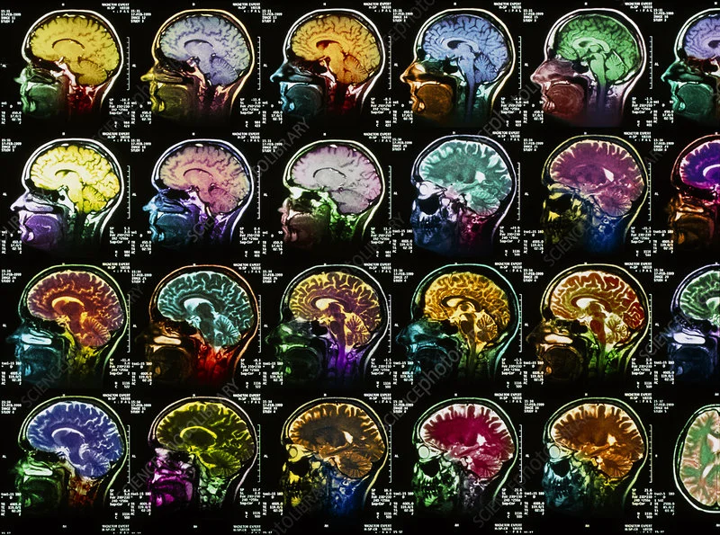
Photon spectral computed tomography: what is it?
Traditional CT scanners use scintillation detectors, which first convert X-rays into visible light and then use photodiodes to convert it into an electrical signal (indirect detection method).
Due to the slow response of the detectors and the low signal-to-noise ratio, information about the energy of individual X-ray photons is lost, so the images obtained can be compared to “black-and-white photographs” — they do not allow for accurate color diagnostics.
Photon spectral CT, on the other hand, uses semiconductor detectors to directly convert X-rays into electrical signals, allowing the energy of each photon to be accurately determined. This makes it possible to visualize the main components of body tissues, such as iodine, calcium, water, and soft tissues, in multispectral mode — effectively creating clearer “color images” of the body, which provides doctors with more visual and accurate diagnostic information.
Advantages of the new technology
The new tomograph provides ultra-high resolution, allowing the smallest pathological structures to be distinguished. In addition, it reduces radiation exposure by 60–70%, and for some organs by up to 80–90%, making the examination much safer for patients, said Du Yanfeng, president of the CT division at United Imaging Healthcare.
“The ultra-high resolution of spectral CT with photon counting allows us to detect lesions earlier and more clearly,” confirmed Yan Fuhua, head of the radiology department at Zhujin Hospital.
According to him, the new tool is especially useful for examining the heart because it is constantly in motion: “This new ‘camera’ can clearly visualize the vessels and structural details of the heart, providing a deeper understanding of the anatomy and functions of the heart.”
Spectral CT of the heart with photon counting
Thanks to its low radiation exposure and high clarity, the new technology will soon become the standard in diagnostics, according to Zeng Mengsu, head of the radiology department at Zhongshan Hospital. The potential applications are enormous: from early diagnosis of cerebrovascular diseases and Alzheimer’s disease (through analysis of the smallest vessels in the brain) to the detection of lung pathologies (such as fibrosis) and small tumors of the liver and pancreas.
Published:
Updated:


Please rate the work of MedTour


