Calls for Ukraine
Calls for Europe
Calls for USA

Nerve damage during surgery is one of the most dangerous complications, which can lead to prolonged recovery, organ dysfunction, and a reduced quality of life for the patient. This is particularly relevant during operations in areas with a high concentration of critical nerve structures, such as neck and head surgery. In situations where even experienced surgeons have to operate “blind” or rely on electrophysiological monitoring, a new approach to nerve visualization could radically change surgical practice.
A recent study published in the journal Nature Communications presented the first-of-its-kind drug bevonesein. This molecule consists of a fluorescent fragment and a short peptide that specifically binds to nerve tissue. After intravenous administration, the drug visualizes nerves as bright greenish-yellow structures in the surgical field, allowing them to be clearly distinguished from surrounding tissues.
During the first and second phases of clinical trials involving 27 patients with neck tumors, the drug’s safety and its ability to improve the visualization of longer nerve segments, which are usually not visible to the naked eye, were confirmed. The study participants required lymph node removal, parotidectomy, and thyroidectomy—procedures during which it is important to preserve the integrity of the cranial nerves. The results of the trials show that the new technology allows unwanted nerve damage to be avoided without the need for additional equipment other than an adapted light source in the microscope.
One of the initiators and co-authors of the study, otolaryngologist Ryan Orosco (University of New Mexico, USA), notes that thanks to more accurate visualization, surgeons can operate faster, safer, and with fewer complications. Orosco himself collaborated with a large team of scientists who created the drug. One of the scientists was biochemist Roger Tsien, who won the Nobel Prize in Chemistry in 2008 for developing green fluorescent protein (GFP), which made it possible to create methods for “labeling” certain molecules and types of tissue, particularly cancerous ones. Orosko is also the coordinator of the current third phase of research, which involves 10 medical centers and is scheduled to be completed in the summer of 2025.
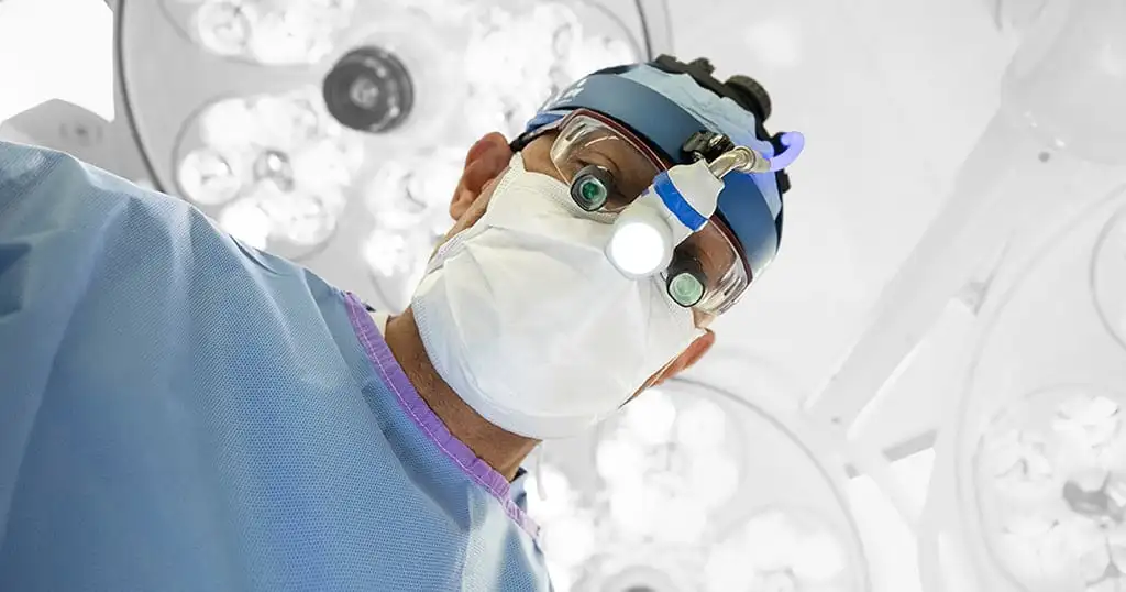
Bevonescein is administered intravenously before surgery and is rapidly excreted by the kidneys, usually within 12 hours. At the same time, its binding to nerve structures remains stable even 6–8 hours after administration, providing a long period of time for high-quality imaging. After the current phase of testing is complete, there are plans to test the use of fluorescent headlamps instead of microscopes, paving the way for the practical and widespread introduction of the technology into routine surgery.
If the study demonstrates clinical efficacy, bevonesein could receive FDA approval, and the drug would then become available for widespread surgical use—not only in head and neck cancer surgery, but also in neurosurgery, maxillofacial, plastic, and even orthopedic practice.
Please rate the work of MedTour
