Calls for Ukraine
Calls for Europe
Calls for USA

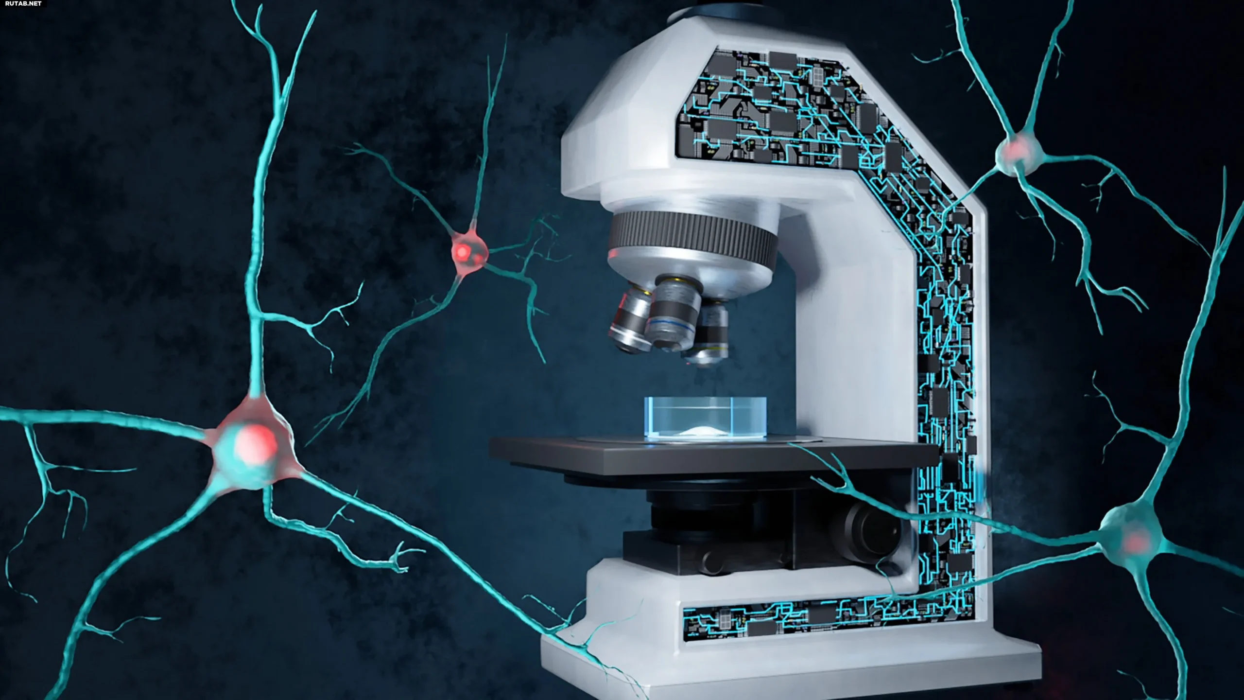
The microscope of the future uses sound to study the brain—its possibilities are endless and astonishing
Researchers at the Massachusetts Institute of Technology (MIT) have made a breakthrough in neurobiology by creating a new generation microscope that uses sound instead of traditional light to image deep regions of the brain. This innovative technology opens up new possibilities for studying brain function and diagnosing neurological diseases.
Problems with traditional microscopy
For decades, microscopy has faced a serious problem: the inability to obtain a clear image of deep layers of the brain without damaging tissue or using special dyes, genes, or chemicals. This has made it particularly difficult to study important areas such as the hippocampus, which is associated with memory and learning.
Three-photon photoacoustic imaging: a revolutionary approach
Scientists at MIT have proposed a fundamentally new approach, combining light and sound to overcome these limitations. The new technology is based on three-photon photoacoustic imaging.
The principle behind this method is as follows:
This method allows images to be obtained without the use of dyes or genetic modifications, which is especially important when working with living tissues, including the brain.
Experiments and results
During the experiments, scientists successfully visualized metabolic activity in cerebral organoids — 3D structures grown from human stem cells. They also successfully tested the method on mouse brain slices up to 0.7 mm thick, demonstrating high resolution.
Particular attention was paid to the NAD(P)H molecule, a key indicator of cellular metabolism and neural activity. This molecule plays an important role in the study of conditions such as Alzheimer’s disease, Rett syndrome, and seizure disorders.
In addition, the method of generating the “third harmonic” (another photonic effect) allows structural images of cells to be obtained without additional labels, i.e., to see the shape and location of cells along with molecular activity, which significantly expands the possibilities of visualization.
Potential applications
By combining several advanced methods — three-photon excitation, label-free imaging, and acoustic detection — researchers have created a platform capable of looking deep into the brain without damaging tissue.
In the long term, this technology may find application in:
The new microscope developed by MIT researchers represents a breakthrough in neuroimaging. This technology opens up new possibilities for studying the brain, diagnosing neurological diseases, and performing neurosurgical operations.
Please rate the work of MedTour
