Calls for Ukraine
Calls for Europe
Calls for USA
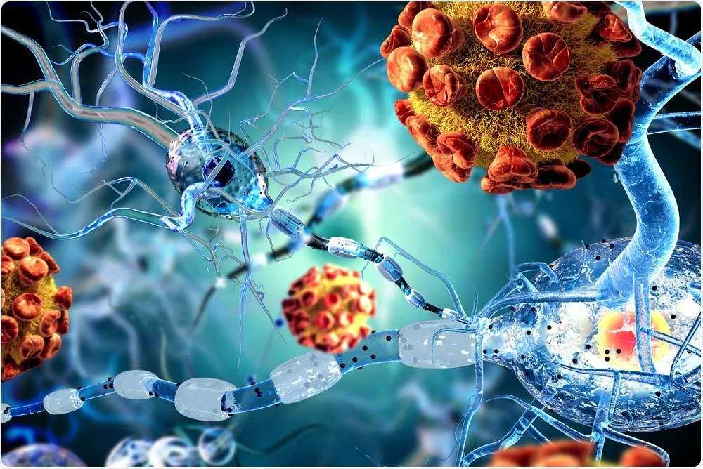
A new study has shown that COVID-19 can lead to the accumulation of proteins similar to those seen in patients with Alzheimer’s disease — not only in the brain, but also in the eyes. This may explain why “brain fog” — a general term for problems with memory and cognitive function — is often mentioned as a symptom of coronavirus infection.
Scientists at Yale University have studied the possible similarities between the neurological effects of COVID-19 and Alzheimer’s disease. They tested whether the SARS-CoV-2 virus is capable of causing the formation of amyloid plaques, which could explain cognitive impairment after the disease.
“There is growing evidence of a link between COVID-19 and ‘brain fog’ — a common symptom after infection,” said senior author Brian Hufler, an ophthalmologist at Yale School of Medicine. “Although the mechanisms behind this phenomenon are not fully understood, scientists have found that the virus can trigger the accumulation of beta-amyloid in the central nervous system.”
The retina is part of the central nervous system, and as one of the most accessible structures for clinical study, it can serve as an indicator not only of vision but also of brain function. It has previously been established that in patients with Alzheimer’s disease, beta-amyloid accumulates in both the brain and the retina, making it a promising target for diagnosis.
For the study, the scientists used postmortem retinal samples from patients, as well as lab-grown retinal organoids—3D models made from stem cells. They analyzed different types of cells by measuring the level of RNA in their nuclei to determine protein production. Particular attention was paid to two proteins—neuropilin-1 (NRP1) and angiotensin-converting enzyme 2 (ACE2)—which SARS-CoV-2 is thought to use to enter neurons.
It turned out that NRP1 is present in neurons and glial cells of the retina in people who have had COVID-19, indicating a possible route of viral entry into the eyes. Patients without dementia but with a history of COVID-19 showed increased beta-amyloid accumulation, reminiscent of changes in Alzheimer’s disease. A similar effect was observed in retinal organoids after exposure to the coronavirus spike protein.
Interestingly, when an NRP1 inhibitor was added, the scientists were able to prevent the increase in beta-amyloid levels in tissues exposed to the virus. This opens up the possibility of developing NRP1-targeted therapies to combat neurological complications of COVID-19, such as “brain fog.”
“The role of NRP1 in beta-amyloid aggregation provides a specific molecular target for further research,” notes Hafler. “Our work has shown that contact with SARS-CoV-2, especially its spike protein, can cause the formation of amyloid clusters in both the human retina and organoids.”
In addition, the study supports the hypothesis that beta-amyloid may have a protective function in the brain. Previously, these plaques were thought to directly cause Alzheimer’s disease, but now they are more often seen as markers of hidden threats. Beta-amyloid is structurally similar to known antimicrobial peptides, and some data point to its role in the brain’s immune defense.
“This supports the antimicrobial hypothesis of Alzheimer’s disease, suggesting that beta-amyloid may be part of the brain’s innate immune response to viral infections,” says Hafler. The study authors note that other viruses are also capable of triggering beta-amyloid accumulation and emphasize the need for further research. Hafler’s team is currently conducting clinical studies to determine whether COVID-19 increases the long-term risk of developing Alzheimer’s disease.
“Our ultimate goal is to prevent the long-term neurological consequences of COVID-19 and to explore NRP1 inhibitors, as well as other modulators of virus-host interactions, as potential therapies for preventing virus-induced amyloid pathology and Alzheimer’s disease,” concludes the scientist.
Published:
Updated:


Please rate the work of MedTour


