Calls for Ukraine
Calls for Europe
Calls for USA
Bone scintigraphy is a method of radiation imaging of bone tissue. It allows to diagnose a number of diseases, including primary or metastatic bone cancer, bone infection (osteomyelitis), inflammation and fractures that may not be visible on traditional x-rays.
Skeletal scintigraphy makes it possible to assess the function of bone tissue and visualize the metabolic processes that occur in it, which is not available for other diagnostic methods, such as x-rays and computed tomography.
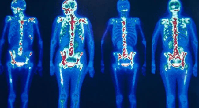
Bone scintigraphy helps detect pathological changes in bones through the use of small amounts of radiopharmaceuticals called radioactive tracers. They are injected into the patient’s bloodstream. The drug passes through the area of study and emits gamma radiation, which is recorded and transmitted to the computer by a special camera. The computer processes the information and creates images of the patient’s bones.
Since bone scintigraphy is able to accurately determine the molecular activity in the tissues of the body, it allows to diagnose the disease at an early stage, much earlier than a conventional x-ray examination. Also, bone metastases can be seen with bone scintigraphy before they can be visualized using other diagnostic techniques.
The study is prescribed to diagnose and monitor the effectiveness of the treatment of diseases such as:
In addition, bone scintigraphy is used to assess the condition of endoprostheses.
Preparation for the study includes several recommendations:
The doctor will give more detailed instructions immediately before the scan.
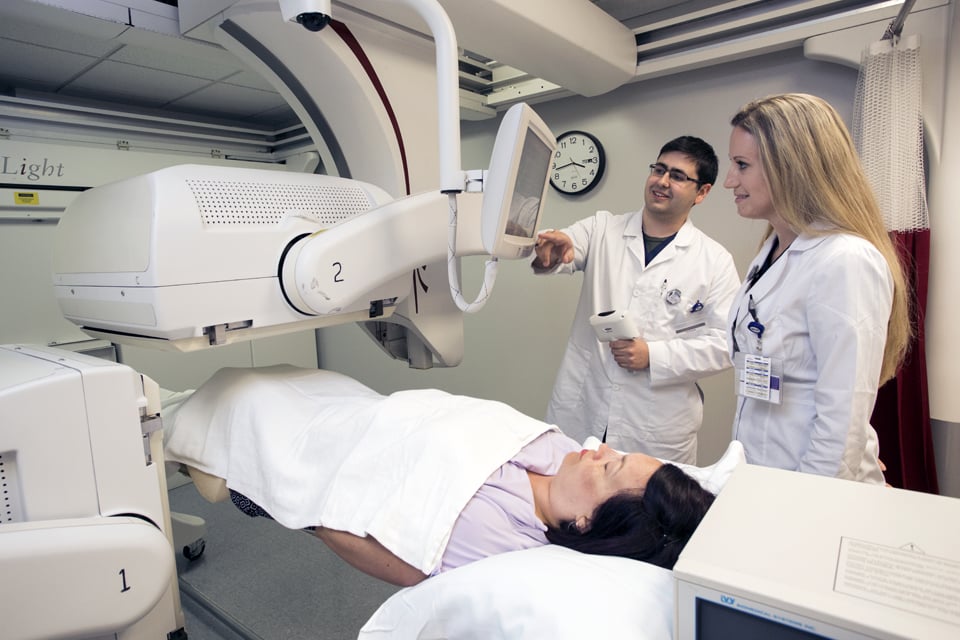
A patient lies on the examination table. A nurse will insert an IV catheter into a vein in your arm. It is necessary for the injection of the radiopharmaceutical. The medicine enters the bloodstream and is absorbed by the bones for some time.
The length of time between the injection and the subsequent scan varies depending on the reason for the study. Sometimes the images are taken immediately after the administration of the radiopharmaceutical, but more often the scan is performed after 2-4 hours, so that the radioactive tracers have time to penetrate into the bone tissue. During this time, the patient will be asked to drink 4 to 6 glasses of water to flush out any residual radioactive material that has not been absorbed by the bones. Before scanning, you need to empty your bladder.
During the scan, the camera can rotate around the patient’s body or remain in one position. In some cases, the patient is asked to change body position between a series of images. While the camera is scanning, you must remain still. The study is painless and is performed with minimal discomfort for the patient.

Sign up for a procedure bone scintigraphy
Fill in your details and our medical coordinator will contact you for a consultation
Pay for the Procedure
Bone scintigraphy may be associated with the following adverse reactions:
Is bone scintigraphy dangerous?
Although radioactive isotopes are used to conduct this study, the exposure of the patient during the study is so small that this technique is suitable for diagnosing children from the first year of life.
For the entire period of clinical use of radiopharmaceuticals in world practice, not a single pronounced allergic reaction has been described. This is due to the fact that the amount of the administered radiopharmaceutical is minimal, as well as the fact that it is biologically inert. The isotopes used for bone scintigraphy are short-lived, they quickly decay, cease to emit radiation and are excreted from the body.
Are there any contraindications for bone scintigraphy?
Absolute contraindications to the study are:
Breastfeeding mothers can continue breastfeeding one day after the procedure is completed.
What happens after the procedure?
After the study, the patient does not pose a danger to others and should not experience any discomfort. However, within 24 hours after bone scintigraphy, close contact with children and pregnant women should be avoided, and the amount of fluid consumed should be increased to 2-2.5 liters.
What are the benefits of bone scintigraphy?
Bone scintigraphy allows doctors to obtain unique information that is not available using other diagnostic methods. This method makes it possible to diagnose primary and metastatic bone cancer at an early stage, osteomyelitis, bone infection, as well as fractures that are not visible on conventional x-rays.
Where is the best hospital to undergo bone scintigraphy?
To make an appointment for a procedure, please contact our coordinating doctors by phone or using the form provided on the website. We will select the clinic and doctor that best suits your problem. We do not charge for our services.
Published:
Updated:


Information on this webpage verified by the medical expert

We are always happy to help you. Write your question by filling out the form. We are happy to answer all your questions.


Please rate the work of MedTour

