
Trophic ulcer. How to cure and accelerate healing
A trophic ulcer, also known as a neurotrophic ulcer, is a type of chronic wound that usually occurs as a result of impaired nerve function or decreased blood supply to a specific area of the body. They usually affect the lower extremities, especially in people with conditions such as diabetes, peripheral neuropathy or vascular
diseases.
What a trophic ulcer looks like. Stages
Trophic ulcers can have different stages, each of which is characterized by specific visual characteristics. The staging system commonly used to describe the progression of trophic ulcers is based on the Wagner classification system. Here is a general description of each stage:
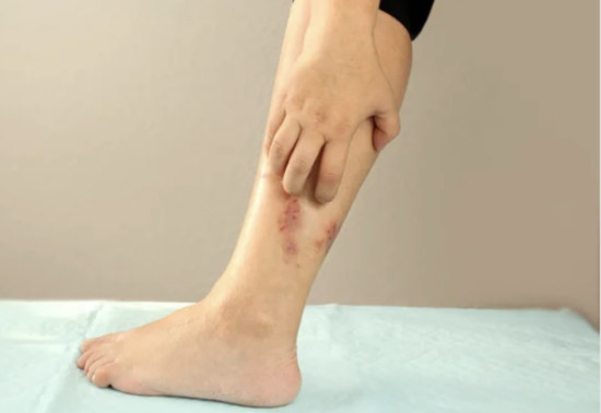
Stage 0: pre-ulcer lesion
- The skin is not damaged, but there are signs of impending ulceration.
- May manifest as areas of redness, discoloration, or calluses.
- The skin may appear warmer or cooler compared to the surrounding areas.
Stage 1: Superficial ulceration
- Ulceration spreads to the epidermis, but does not reach deeper tissues.
- The ulcer looks like a shallow open wound.
- May have a red or pink wound bed.
Stage 2: The ulcer spreads to the tendon, bone or capsule of the joint.
- The ulcer penetrates deeper into the subcutaneous tissue and can reach tendons, bones or joint bags.
- The wound may be deeper with visible tissue loss.
- May have a yellow or necrotic (dead tissue) wound bed.
Stage 3: deep ulcer with abscess or osteomyelitis.
- The ulcer spreads to deep structures such as abscesses (pus-filled pockets), or affects the underlying bone, leading to osteomyelitis (bone infection).
- The wound may have significant tissue loss with exposed bone or deep cavities. There are often signs of infection, including redness, swelling and purulent discharge.
Stage 4: Gangrene or gangrenous ulceration.
- Ulceration is extensive, often with extensive tissue death (gangrene).
- The area may appear black or necrotic due to lack of blood supply.
- Associated with severe infection and tissue destruction.
It is important to note that Wagner`s classification system is primarily focused on diabetic foot ulcers, but can also be applied to other types. Proper diagnosis and stage determination should be performed by a medical professional with
experience in wound care to determine an appropriate treatment plan based on the specific characteristics and underlying causes of the ulcer.
Causes of trophic ulcers
Trophic ulcers can be caused by various factors, and the underlying cause may vary depending on the patient and the specific circumstances. In most cases, they arise due to such factors:
- Pressure or friction. Prolonged pressure or friction on a certain area of the body can lead to the development of trophic ulcers. This often occurs in people who are bedridden, wheelchair bound or have limited mobility, which leads to constant pressure on the bone protrusions.
- Peripheral neuropathy — nerve damage or dysfunction, especially peripheral neuropathy, can contribute to the formation of trophic ulcers. Conditions such as diabetes, peripheral neuropathy, or spinal cord injuries can impair sensitivity and reduce the ability to perceive pain or discomfort, increasing the risk of tissue
damage and ulceration.
- Vascular insufficiency. Poor blood circulation and insufficient supply of oxygen and nutrients to tissues can lead to trophic ulcers. Vascular diseases such as peripheral artery disease (PCA), venous insufficiency, or conditions that cause narrowing or blockage of blood vessels can disrupt tissue health and lead to ulcers.
- Injury. Direct trauma, such as cuts, burns, or other forms of bodily injury, can cause damage to the skin and underlying tissues. If the affected area does not heal properly or cannot heal due to impaired blood flow or neuropathy, a trophic ulcer may develop.
- Infections. Infections of the skin or subcutaneous fat can contribute to the development of trophic ulcers. Infection can disrupt the healing process and lead to tissue damage, splitting or necrosis.
- Autoimmune diseases. Some autoimmune diseases, such as systemic lupus erythematosus (SLE) or vasculitis, can cause inflammation and damage to blood vessels and tissues. This can contribute to the development of trophic ulcers.
It is important to note that in many cases trophic ulcers occur as a result of a combination of factors rather than a single cause. There are factors that can increase their risk, including poor nutrition, weakened immune function, obesity, old age and some medications that affect wound healing or increase the predisposition to ulcers. Proper diagnosis and identification of the root cause are necessary to develop an effective treatment plan and prevent recurrence of trophic ulcers.
Symptoms of trophic ulcers
Trophic ulcers can manifest various symptoms, and the specific symptoms may depend on the underlying cause and severity of the ulcer. Here are some common symptoms associated with trophic ulcers:
- Open wound: Trophic ulcers usually look like open sores or wounds on the skin. The size, depth and appearance of the ulcer may vary depending on the stage and severity of the condition.
- Slow healing. Trophic ulcers are chronic wounds that tend to heal slowly or may not heal without appropriate treatment. The lack of healing progress over time is a characteristic feature of trophic ulcers.
- Pain or discomfort. Some people with trophic ulcers may experience pain, soreness or discomfort in the affected area. The level of pain can vary from mild to severe, depending on the individual tolerance of pain and the main involvement of the nerve.
- Redness and inflammation. The skin around it may be red, inflamed or irritated. Inflammatory changes may be a sign of infection or tissue damage.
- Drainage or exudate. Trophic ulcers can produce various types of drainage or exudate, including clear fluid, serous fluid, pus or blood. The type and amount of discharge may indicate the stage and severity of the ulcer.
- Smell. In some cases, trophic ulcers may have an unpleasant odor, especially in the presence of infection or necrotic (necrotic) fabrics. A fetid odor can be a sign of bacterial colonization or tissue destruction.
- Skin texture changes. The texture of the skin surrounding the trophic ulcer may vary. It can become hardened, thickened or calloused due to chronic pressure or tissue damage.
- Numbness or change in sensitivity. If trophic ulcers are associated with peripheral neuropathy, people may experience numbness, tingling or loss of sensitivity in the affected area. This can make it difficult to detect pain or discomfort and delay seeking appropriate help.
Diagnosis of trophic ulcers
The diagnosis, as a rule, requires a thorough assessment by a medical professional, it is desirable that this doctor be an experienced surgeon. The diagnostic process may include the following steps:
- Medical history. The doctor collects an exhaustive medical history, including any underlying conditions or factors that may contribute to the development of trophic ulcers. Information about previous injuries, operations or appropriate treatment will also be taken into account.
- Physical examination: A thorough physical examination is mandatory to assess the ulcer and surrounding tissues. The doctor will check the size, depth, appearance and characteristics of the ulcer, as well as any signs of infection or complications. It can also assess the surrounding skin, blood circulation and nerve function.
- Direct assessment of the wound. Trophic ulcers are assessed by various parameters, including size, depth, presence of necrotic tissues, exudate, signs of infection and tissue viability. This assessment helps to determine the severity and stage of the ulcer, which determines the approach to treatment.
- Imaging studies: in some cases to assess blood flow, identify any hidden vascular abnormalities or obstruction, and evaluate Imaging studies, such as ultrasound Dopplerography or angiography, may be recommended for the condition of blood vessels supplying the affected area.
- Laboratory tests: Depending on the clinical situation, laboratory tests may be conducted to assess general health, exclude background infections, assess blood glucose levels (in the case of diabetes) and check other potential contributing factors.
- Biopsy. In certain situations, a biopsy may be performed to obtain a small sample of tissue for further examination under a microscope. A biopsy can help identify specific causes or underlying conditions that contribute to the occurrence of trophic ulcers.
The diagnosis includes consideration of both the clinical picture and the main factors contributing to the development of ulcers. This comprehensive assessment helps to determine the appropriate treatment plan and management strategies to accelerate healing and prevent complications.
How to treat a trophic ulcer?
Treatment involves a comprehensive approach aimed at accelerating healing, preventing complications and eliminating the root cause. The specific treatment plan may vary depending on the characteristics of the ulcer and the overall health of the person. It is very important to consult with an experienced doctor who has experience working in a specialized center to make an accurate diagnosis and individual treatment recommendations. However, some approaches to treatment are mandatory and common:
- Wound care and bandages. Proper wound care is crucial for the treatment of trophic ulcers. It includes regular cleaning of the ulcer with a mild antiseptic solution or sterile saline solution to prevent infection. Depending on the characteristics of the wound, appropriate dressings can be used to create a moist wound environment, absorb excess exudate, accelerate healing and protect the ulcer from external contamination.
- Pressure relief — reducing pressure on the affected area is vital for the healing of a trophic ulcer. This can be achieved by using special pillows, mattresses or pads that redistribute pressure and relieve the load on the ulcerated area. It is also important to change position frequently and avoid prolonged periods of
immobility.
- Unloading and elevation: If a trophic ulcer is located on the foot or lower leg, unloading this area is crucial to reduce pressure. This may include the use of assistive devices such as crutches, walkers or wheelchairs to avoid loading the affected limb. Lifting the affected limb while resting can help reduce swelling and
improve blood circulation.
- Treatment of infection. If there are signs of infection, appropriate measures should be taken to control and treat the infection. To do this, antibiotics are used, which are either injected directly into the wound or applied systematically.
- Surgical treatment. In cases where non-viable or necrotic tissues are present in the ulcer, sanitation may be required. Sanitation helps to remove dead tissue and create a healthier wound bed for healing. Sanitation can be achieved using various methods, such as surgical treatment, enzymatic treatment (rarely used) or autolytic
treatment (using a special gel for wound treatment).
- Compression therapy. In some cases, compression therapy may be useful, especially for trophic ulcers associated with venous insufficiency. Compression bandages or stockings help to improve blood circulation and reduce swelling.
- Nutritional support: Adequate nutrition plays an important role in wound healing. It is necessary to eat a balanced diet rich in proteins, vitamins and minerals. In some cases, a nutritionist may provide guidance on specific dietary recommendations or dietary supplements to support healing. MedTour recommends Dr. Alexander Kirilenko.
- Treatment of concomitant diseases. Treatment of conditions that contribute to the occurrence of trophic ulcers is important for long-term healing and prevention of relapses. Such methods include blood glucose control in diabetes, treatment of vascular diseases or treatment of other diseases.
The latest methods of treatment of trophic ulcers
- The use of stem cells in the treatment of trophic ulcers is an area of active research. Although stem cell therapy for trophic ulcers has not yet entered the treatment standards, it has managed to show impressive results. Stem cells have a unique ability to differentiate into different types of cells and have regenerative properties. They can be obtained from a variety of sources, including adipose tissue (fat), bone marrow or umbilical cord tissue.
Stem cells in the treatment of trophic ulcers can be used in several ways:
- Stem cell transplantation: Stem cells can be isolated and prepared for transplantation to the site of the lesion. These cells can promote tissue regeneration, angiogenesis (formation of new blood vessels) and reduce theinflammatory response. Transplanted stem cells can also release growth factors and
cytokines that promote the healing process.
- Products derived from stem cells. Instead of direct stem cell transplantation, it is possible to use products derived from stem cells, such as growth factors, cytokines or extracellular vesicles. These products contain useful bioactive molecules secreted by stem cells, and they can be applied topically or injected into an ulcer to
stimulate healing processes.
- Combination therapy. Stem cell therapy can be used in combination with other known treatments. For example, stem cell therapy can be used in combination with appropriate wound care, unloading or compression therapy to improve the overall healing process.
How is the treatment of trophic ulcers using stem cells?
Treatment with stem cells takes place in several stages.
1 Stem cell collection: Stem cells can be obtained from a variety of sources, including adipose tissue (fat), bone marrow or umbilical cord tissue. The chosen source will depend on factors such as availability, availability and the specific protocols of the treatment used.
2 Isolation and preparation of stem cells: After collection, stem cells are isolated from a tissue sample and processed to produce a concentrated population of stem cells. For highlighting and cleaning Various methods can be used, such as enzymatic cleavage or cell centrifugation.
3 Stem cell delivery: Stem cells can be delivered to the site of a trophic ulcer in various ways, depending on the specific treatment approach. These methods may include:
- Local injection. In this case, stem cells are injected directly into the ulcer or into surrounding tissues. This allows you to purposefully deliver stem cells to the right place.
- Topical application: Products derived from stem cells, such as growth factors or cytokines, can be applied topically to the ulcer. These products contain beneficial bioactive molecules secreted by stem cells that can promote tissue healing and regeneration.
- Application to scaffolds: Stem cells can be combined with biomaterial scaffolds that act as a supporting structure for cells. These scaffolds can be placed on the bedof the ulcer, facilitating the interaction between stem cells and the wound environment.
4 Follow-up and monitoring. After stem cell therapy, careful monitoring and follow-up is necessary to assess the progression of the ulcer and the response to treatment. This may include regular assessment of the wound, assessment of healing parameters, and adjustment of the treatment plan as needed.
Prevention of trophic ulcers
Prevention involves eliminating the root causes and taking measures to minimize the risk of their development. Here are some general strategies for the prevention of trophic ulcers:
- Skin care. Proper skin care is necessary to maintain its health and integrity. Keep your skin clean and hydrated to prevent dryness and cracking. Use mild soap and avoid excessive use of hot water. Regularly inspect the skin for signs of redness, irritation or injury.
- Maximum pressure relief: Reduce pressure and friction on vulnerable areas of the body by practicing regular position changes and using appropriate pads or pillows. This is especially important for people who are sedentary or spend a long time in a sitting or lying position. Use specialized devices or auxiliary means for pressure relief in accordance with the recommendations of medical professionals.
- Unloading: If you have a condition that increases the risk of trophic ulcers, such as peripheral neuropathy or venous insufficiency, it is important to unload sensitive areas. This can be achieved with the help of auxiliary devices such as crutches, walkers or wheelchairs, or with the help of special pillows or mattresses.
- Maintaining a healthy weight. Being overweight can contribute to increased pressure on the feet and lower extremities. Maintaining a healthy weight can help reduce the risk of developing trophic ulcers associated with excessive pressure.
- Regular physical exercises. Regular exercise improves blood circulation and
overall health.
- Foot care for diabetes. Patients with diabetes need to pay special attention to foot care. It is necessary to examine your feet daily for any signs of cuts, blisters or ulcers, as well as keep your feet clean and moist, trim your nails regularly and wear suitable shoes.
Our best specialist doctors
The MedTour team has extensive experience in providing medical care to different groups of patients, including those with trophic ulcers. For such clinical situations, we recommend Dr. Alexander Kovalchuk if the patient is in Ukraine and Dr. Ivan Badin for patients outside of Ukraine.
Coordinating doctors will answer any question about the treatment of trophic ulcers with stem cells absolutely free of charge.
F.A.Q.
1. What is the cost of treating trophic ulcers with stem cells?
The cost of treating trophic ulcers with stem cells depends on several factors, including the country or region where the treatment is offered, the specific stem cell therapy protocol used, the source of stem cells (for example, autologous or allogeneic), the number of procedures required and additional related medical services. This ranges from several hundred to several thousand dollars per course.
2. How many courses of stem cells are required to achieve an effect in the treatment of trophic ulcers?
This issue is solved individually based on the clinical situation and the patient`s state of health, but most often patients need 3-4 courses.
3. How long does it take for a noticeable clinical effect to occur in trophic ulcers with stem cells?
It usually takes 1-3 weeks for the first signs of improvement to appear, and up to 3 months to achieve full effect. But this is also an average indicator, only the attending physician can predict the condition of a particular patient.
