-
 News
When glucose levels are low, chemotherapy ceases to affect cancer cells
-
 News
Excessive treatment of prostate cancer in older men may reduce quality of life without increasing its duration
-
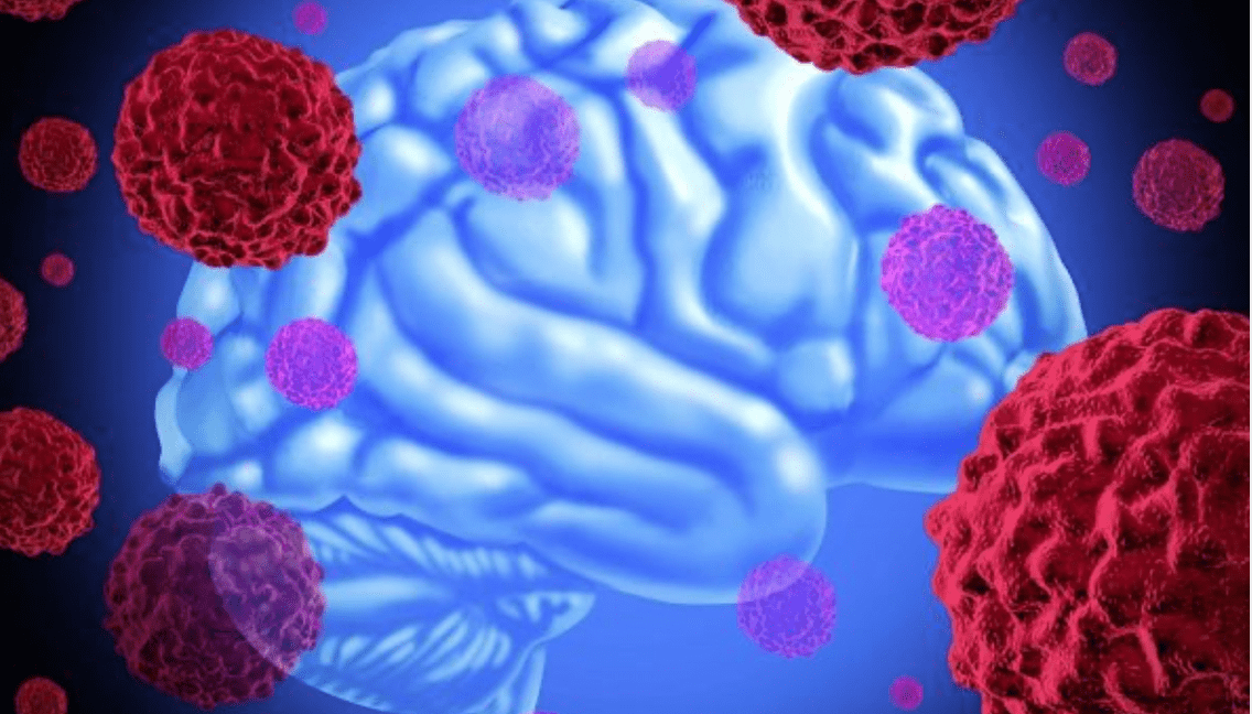 News
Brain cancer can be cured by viruses
-
 News
Ways to reduce lymphatic pain in breast cancer have been found
-
 News
Scientists have turned bacteria into a powerful weapon against cancer
All news
Cerebral artery aneurysm treatment
A cerebral artery aneurysm is a pathological bulge of a cerebral artery in the shape of a balloon. The type of aneurysm is determined using the diameter of the vessel: less than 2 mm — microaneurysms, 3-15 mm — normal size, 16-24 mm — large and over 25 mm — giant aneurysms.
- Half a million fatal aneurysm ruptures occur each year;
- Disease occurs at the ages of 35-60 years.
Diagnostic methods: CT, MRI, cerebral angiography, lumbar puncture.
Treatment: endovascular embolization, clipping, vessel wall wrapping, balloon angioplasty, treatment with new generation drugs.
MedTour patients recommend clinics for the treatment of cerebral artery aneurysm:
Doctors for the treatment of cerebral artery aneurysm
Patient reviews
Frequently Asked Questions
It is a ball-like bulge on the wall of an artery in the brain. It occurs where the blood vessel wall is weakened. This tissue weakness can be congenital or acquired.
Symptoms are due to increased intracranial pressure:
- Sudden severe headache,
- nausea (does not go away even after vomiting),
- a feeling of stiffness in the neck,
- drowsiness,
- loss of consciousness or coma.
- Congenital vascular weakness,
- high pressure,
- arteriosclerosis,
- smoking,
- presence of other diseases (influenza),
- foci of pus in the body.
The type of aneurysm depends on the number of layers of artery tissue involved. Aneurysms can vary in size. The more the vessel wall protrudes, the greater the risk of tissue rupture.
Blood passes between the middle of the three meninges and the surface of the brain — subarachnoid hemorrhage occurs. Symptoms of a rupture: a sharp headache, a feeling of nausea (does not go away even after vomiting), impairment or loss of consciousness. It is a life-threatening form of stroke and must be treated immediately with surgery.
The gap can cause changes in speech, vision, or movement. The consequences depend on which part of the brain is affected and how serious the circulatory disorder is.
Often, a cerebral aneurysm is found incidentally by CT, MRI, or cerebral angiography. If an aneurysm rupture is suspected, cerebrospinal fluid is examined.
Symptoms are corrected with medications, including pain relievers and antihypertensive drugs. In a severe case or if the artery is ruptured, it is necessary to strengthen it surgically or remove the aneurysm (by clipping or wrapping).
Estimated prices:
- Consultation with a neurologist from $ 100,
- MRI from $ 500,
- CT scan from $ 400,
- Angiography from 1300 $,
- Surgical treatment from $ 10,000.
Do you want to know the exact price or find out the price differences in clinics? Leave a request on the platform MedTour and the Doctor-Coordinator will consult you for free!
International methods of diagnosis and treatment of cerebral artery aneurysm
Vascular dilatation or rupture of an aneurysm is detected by magnetic resonance imaging (MRI) or computed tomography (CT). When an aneurysm ruptures, the doctor has enough symptoms to suspect a diagnosis. Other diagnostic tests include:
- examination of cerebrospinal fluid,
- cerebral angiography.
Visualization techniques
If a ruptured aneurysm in the brain is suspected, computed tomography (CT) is done. With this X-ray study, blood flow in the brain, hemorrhage and ruptured aneurysm can be clearly observed.
Magnetic resonance imaging (MRI) does not use X-rays and provides more information about bleeding and cerebral vasodilation than CT.
Laboratory methods
In patients with a serious condition, fluid in the brain and spinal cord (CSF) is examined. If the aneurysm ruptures, there will be red blood cells in the cerebrospinal fluid. A special needle is used to draw fluid from the spinal canal. This procedure is called lumbar puncture.
Cerebral angiography
Cerebral angiography is used to establish a definitive diagnosis. From the groin to the brain, a thin, flexible catheter is passed through a large artery. The arteries of the brain are stained with a dye and a ruptured aneurysm is clearly visible on CT.
Methods for the treatment of cerebral artery aneurysm
The first step is to assess the presence of symptoms and risk factors. In the absence of symptoms and risks, it is sufficient to undergo regular examination. But if there is a risk of rupture, it is necessary to strengthen the artery or clamp the aneurysm.
Treatment options depend on:
- the age of the person,
- the severity of symptoms,
- location of the aneurysm,
- the size of the aneurysm,
- forms of the vasculature of the brain,
- presence of other diseases.
Endovascular method
In endovascular embolization, a catheter is used to advance a fine wire mesh from the groin into a weakened artery in the brain. This stent strengthens the vessel wall from the inside. Then the micro-spiral platinum spiral fills the defect in the vessel wall.
Neurosurgical methods
These methods are used when it is impossible to perform the endovascular method or when urgent treatment is required.
The surgeon opens the skull and chooses one of the options for eliminating the problem:
- clamp the weakened area of the artery with a titanium clamp (clipping),
- strengthen the vascular wall from the outside in the defective place (wrapping),
- Introduce balloons before and after a cerebral aneurysm (balloon angioplasty).
Drug treatment
Symptoms and complications are usually treated with medications. Pain relievers for headaches and calcium channel blockers are prescribed to prevent vasospasm. Narrowing of the intracranial arteries can lead to severe and fatal cerebral infarctions.
How to choose a clinic for treatment abroad
- availability of special equipment for various neurosurgical and endovascular treatment methods;
- modern CT and MRI machines;
- experienced specialists (neurologists, neurosurgeons, nurses);
- use of international diagnostic and treatment protocols.
Published:
Updated:







Велика подяка за життя. Возняк Олександр Михайлович, Зенкевич Ярослав Павлович.
Кажуть Феофанія для міністрів, ні ця лікарня для людей. І я цьому приклад. Кожного року після операції присилаю МРТ на контроль. Ні разу не відмовили. Всіх пам’ятають, хоч я простий український роботяга. Ці люди від Бога, для людей!