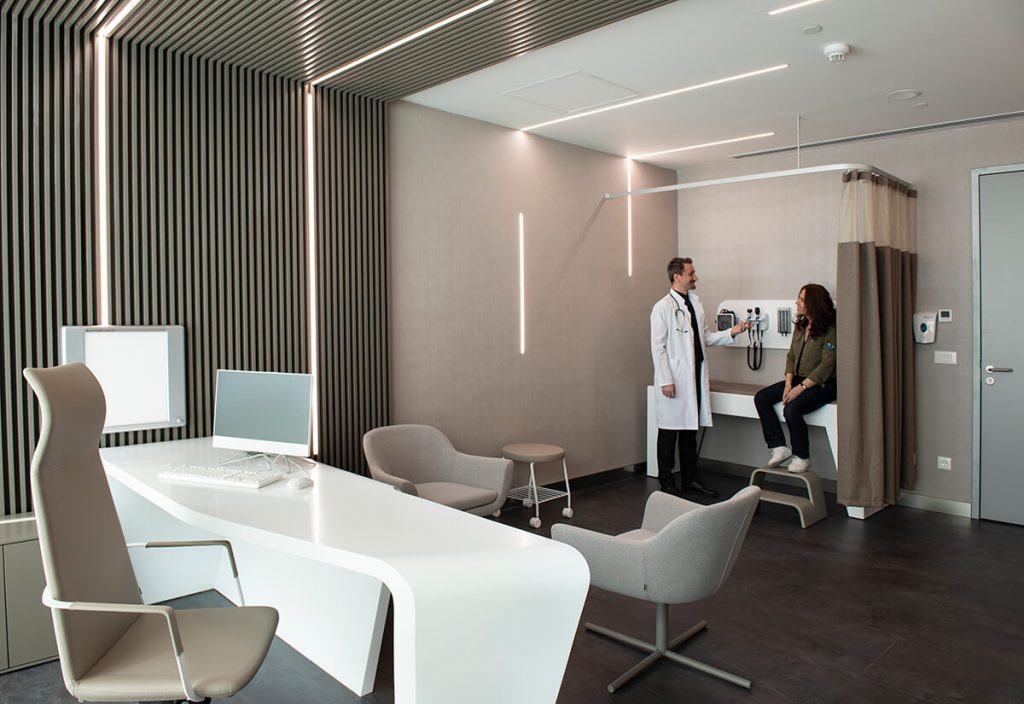
Features of Liv Hospital Vadistanbul
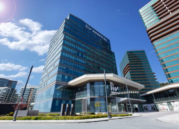
Liv Hospital Vadi Istanbul was established in 2021 and built according to modern standards. Despite the fact that the clinic is located within the city, it is surrounded by greenery. There are parks and forest areas around the hospital building, where patients have the opportunity to walk and enjoy fresh air.
Liv Vadi Istanbul has more than 90 specialized departments, including cardiology, oncology, orthopedics, neurology, pediatric and adult surgery, as well as sports medicine department. The hospital has 302 beds and 14 operating rooms equipped with modern and advanced equipment. Robotic and traditional surgeries are performed in the clinic in the fields of cardiac surgery, neurosurgery, orthopedics and traumatology, as well as general, gynecological, and urological surgery.
Liv Hospital Vadi Istanbul also actively collaborates with Boston Children’s Hospital, allowing patients to receive a second opinion on complex cases involving the diagnosis and treatment of children. This underlines the hospital’s commitment to providing high-quality medical care.
Robotic Surgery at Liv Hospital Vadistanbul
The Robotic Surgery Center at Liv Hospital Vadistanbul in Turkey offers a wide range of high-precision surgeries on the gallbladder, pancreas, lungs, liver, stomach, intestines, and other organs. These are minimally invasive surgical interventions that do not require large incisions. This approach minimizes the risk of bleeding and infection, as well as reduces the rehabilitation period for patients.
Another advantage of robot-assisted surgeries is their high precision. During the procedure, surgeons control the mechanized “hands” of the robot from a specialized console. They control the process using a detailed, highly magnified image of the surgical field. The robot’s manipulators can rotate 360 degrees, which allows the surgeon to perform precise and smooth movements that are not possible with traditional laparoscopy. The robotic system also eliminates hand tremors, minimizing the “human factor” and the possibility of medical error.
At Liv Hospital Vadi Istanbul, robotic-assisted surgeries are performed in the following areas:
General surgery:
- esophageal cancer;
- stomach cancer;
- adrenal cancer;
- pancreatic cancer;
- liver cancer;
- colorectal cancer.
Gynecological surgery:
- cervical cancer;
- uterine cancer;
- ovarian cancer.
Urological surgery:
- prostate cancer;
- kidney cancer;
- bladder cancer.
Robotic heart surgery
Advancements in cardiac surgery, particularly over the last 15 years, have allowed for the expansion of the use of minimally invasive techniques as an alternative to the traditional approaches. In particular, robot-assisted coronary artery bypass surgery offers significant benefits for patients in terms of safety and recovery time.
At Liv Hospital Vad Iistanbul, robotic coronary artery bypass surgery is performed through three small incisions in the armpit and one 4-centimeter incision in the rib area. The procedure is performed on a beating heart, without stopping it. A camera inserted through one of the incisions provides the surgeon with a clear, three-dimensional view of the surgical field, magnified 8-10 times.
Since the chest does not need to be opened, patients can return to their normal lives much faster after robotic coronary artery bypass surgery. Additionally, the risks of bleeding and infection are minimized, and the procedure leaves no noticeable scars.
Robotic Bariatric Surgery in Turkey
Bariatric and metabolic surgery has proven highly effective in treating obesity and related conditions, such as diabetes and high blood pressure. Currently, Liv Hospital Vadi Istanbul offers such surgeries using robotic systems.
Gastric bypass is considered the “gold standard” of bariatric and metabolic surgery. The use of a robotic system allows the surgeon to clearly visualize the surgical field and operate in limited and hard-to-reach places, minimizing surgical trauma.
One of the most serious complications of gastric bypass and other bariatric surgeries is the leakage of stomach or intestinal contents into the abdominal cavity. The robotic approach significantly reduces the likelihood of this complication compared to traditional laparoscopy.
Brachytherapy at Liv Hospital Vadi Istanbul in Turkey
Brachytherapy is a treatment method in which radioactive sources are delivered close to the tumor. This method is successfully used to treat gynecological diseases (uterine, cervical, and vaginal cancer), lung cancer, and skin cancer. Today, thanks to the use of visualization methods, such as computed tomography and magnetic resonance imaging, brachytherapy can be used to treat more than 30 types of cancer.
Three-dimensional brachytherapy (3D brachytherapy) for gynecological cancer
Brachytherapy is a form of radiotherapy in which radioactive sources are placed in close proximity to the tumor, making it an essential component in the treatment of gynecologic oncology. This method is most commonly used to treat uterine (endometrial) cancer, cervical cancer, and vaginal cancer. Brachytherapy can be performed as a stand-alone procedure after surgical intervention or as an adjunct to external beam radiation therapy, particularly for patients who are not candidates for surgery.
In recent years, brachytherapy has evolved from two-dimensional to three-dimensional methods, which has become possible thanks to the precise data obtained using computed tomography (CT) and magnetic resonance imaging (MRI). The use of 3D brachytherapy significantly improves treatment outcomes, providing a more targeted effect on the tumor and reliable protection of healthy tissues (bladder, rectum, sigmoid colon and other organs). This approach reduces the risk of side effects, while maintaining high efficiency of therapy.
Skin Brachytherapy (Leipzig Applicator)
Skin brachytherapy using the Leipzig applicator is effectively employed in the treatment of early stages of squamous cell and basal cell carcinoma, particularly for tumors with optimal size and depth. This method helps minimize cosmetic defects, which are often associated with the surgical removal of tumors located on the face.
3D brachytherapy for lung (bronchial) cancer
For patients with lung cancer who cannot undergo external beam radiation therapy, brachytherapy, which involves placing catheters into the main bronchi and airways, may be used.
MR-Linac Radiotherapy at Liv Hospital Vadi Istanbul
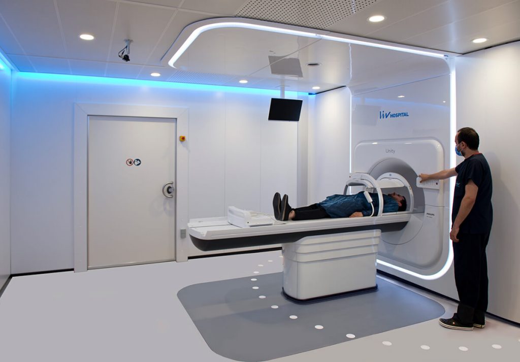
The MR-Linac Unity device, equipped with high-precision imaging capabilities, allows for clear differentiation between tumors and healthy tissues, which is crucial for planning and administering radiation therapy. With real-time imaging, MR-Linac Unity enables precise assessment of the tumor and surrounding tissues throughout the entire treatment process.
This method is effective in treating tumors that move with breathing, such as those located in the lungs or upper abdomen, as well as tumors located near radiosensitive organs such as stomach, kidneys, heart, or spinal cord. MR-Linac Unity enables detailed visualization of soft tissues, clearly separating the tumor from normal tissue, blood vessels, and bone structures, which allows for minimizing radiation exposure to healthy tissue and ensures targeted therapy for the tumor.
